
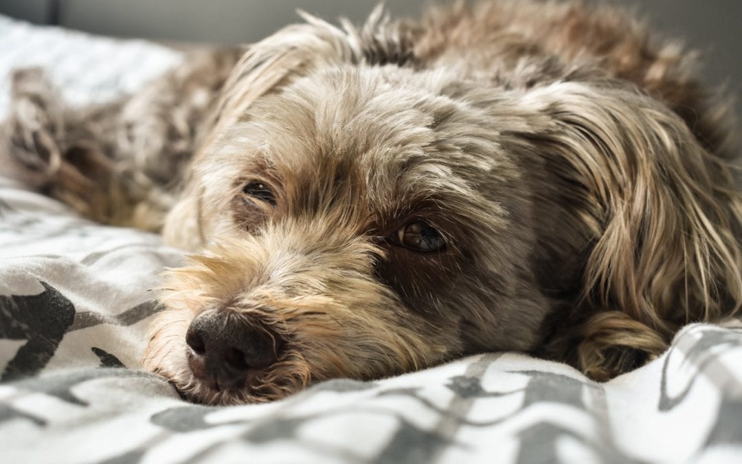
Cataracts, defined as an opacity of the lens, are common in dogs and may result in substantial vision impairment in advanced stages. The most common causes are genetic influences, diabetes mellitus, and age-related factors.
Although cloudiness and vision loss are often the most striking features of cataracts, cataracts are nearly always associated with uveitis (intraocular inflammation). Clinical signs of uveitis are often difficult to recognize for pet owners and primary care veterinarians. However, if left untreated, uveitis may result in pain, intraocular adhesions, lens instability, glaucoma, and retinal detachment. With this in mind, most dogs diagnosed with significant cataracts would benefit from medication to help prevent or control cataract-associated uveitis.

Phacoemulsification for cataract extraction with subsequent placement of an artificial lens is the most effective treatment for improving vision in eyes with advanced cataracts. The technique used in dogs is similar to that in people, and the success rate is high (~80-90%). Not only is vision restored, but the risk of the aforementioned sequelae, like uveitis and glaucoma, may be reduced. However, some of the problems that can occur due to cataracts may also occur after surgery, resulting in vision loss. For best results, careful planning and diligent post-operative care are recommended.
Pre-operative Planning
Several pre-operative steps are necessary for planning before cataract surgery.
- Comprehensive ophthalmic examination to identify and address confounding diseases that may influence the approach or outcome of surgery. In most cases, the veterinary ophthalmologist would also take this opportunity to discuss the details of the surgery with you.

- Because cataracts are often substantial at first presentation to a specialist, veterinary ophthalmologists may be unable to properly evaluate the ocular fundus (i.e., retina and optic nerve) with standard examination equipment. Consequently, electroretinography (ERG) and ultrasonography of the eye(s) may be performed to ensure proper retinal function and vitreoretinal anatomy prior to surgery.
- Gonioscopy to evaluate the iridocorneal (“drainage”) angle is also sometimes performed at the discretion of the ophthalmologist to assess for the risk of post-operative glaucoma. This may be particularly important in breeds commonly affected by glaucoma, like Cocker Spaniels and Labrador Retrievers.
- As is expected in most patients undergoing elective surgery, laboratory work is often recommended prior to undergoing general anesthesia to help ensure appropriate systemic health and guide anesthetic drug selection.
Overall, the above tests may or may not require a second visit to the ophthalmologist, and pet owners should be prepared for the associated fees prior to being approved for cataract surgery.
Post-operative Management
There are 4 challenges of post-operative management that pet owners should consider.
- Patients require oral and topical medications, both antibiotics and antiinflammatories, following surgery. Oral medications are typically administered 1-2 times daily for 2-3 weeks, while the topical medication regimen varies between patients and clinicians. Most patients start out with 3-5 topical medications applied 3-4 times daily, with a gradual reduction in frequency and number of medications over 2-3 months.
- During the medication tapering period, numerous follow-up evaluations are performed. Most patients are evaluated 1 week after surgery, then every 2-4 weeks for several months.
- An E-collar must be worn for at least 2-3 weeks after surgery to prevent rubbing at the eyes while the corneal surgical incisions heal.
- Activity should be restricted for 1 month after surgery with no running, jumping, or rough playing. A harness (rather than a neck lead) is often recommended to limit pressure on the neck during controlled leash walks.
If you’re interested in cataract surgery for your dog (or cat), please reach out to us, and we’d love to schedule a consultation to discuss options and prognosis for your pet!
Before